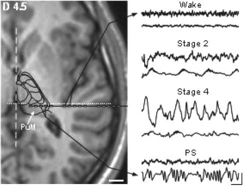
Figure 1.
right: examples of raw bipolar recordings performed simultaneously in the thalamus and cortex during 4 different vigilance states. Note the peculiar delta oscillation appearing in the thalamus during PS. Scale: 250 μV; 1 s. Left: localisation of recording electrode contacts within the lateral part of PuM and the most posterior extent of the insular cortex. Stereotactic horizontal MRI slice is located 4.5 mm dorsal to the horizontal anterior-posterior commissural plane. Localization of thalamic nuclei is based on the fitting of the corresponding sketch of the human thalamic atlas of Morel et al. (1997) with this MRI section. Broken and dotted lines indicate interhemispheric and vertical posterior commissure planes, respectively. Scale: 10 mm.

