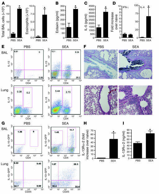
Figure 1. Th2-polarized SEA-induced airway inflammation.
Animals were sensitized with 10 μg SEA on days 1 and 14, followed by 2 intratracheal airway challenges with 10 μg SEA on days 28 and 31. Animals were euthanized 24 hours after the final airway challenge. *P < 0.05, Mann-Whitney U test. (A) Total airway infiltrates and airway eosinophilia recovered in BAL. (B) BAL fluid eotaxin levels measured by ELISA. (C) BAL fluid IL-5 levels measured by ELISA. (D) Lung tissue mRNA, IFNγ (Th1) and IL-4 (Th2). (E) BAL and lung cells stimulated with PMA and ionomycin with brefeldin A for 3 hours and then stained with anti-CD4 (APC) and anti-IL-13 (PE) before acquisition and analysis by FACS. (F) Five-micrometer sections were cut from paraffin-embedded lung tissue and stained with H&E (lower panels) to show cellular infiltration or alcian blue–PAS (upper panels) showing mucin+ goblet cells lining the airways. The mucin response to SEA was 7+, while the inflammatory response to PBS was 2+ and to SEA, 4+. Five lungs from each group were scored in a blinded fashion; original magnification, ×100. (G) Lung cells and BAL recoveries from C57BL/6 IL-10–GFP mice were stained with anti-CD4 (APC) and anti-CD25 (PE) for lung cells, with GFP expression observed in the FL1 channel. (H) RNA was extracted from lung tissue, with IL-13Rα2 mRNA quantified by quantitative reverse transcription PCR (qRT-PCR). (I) Soluble IL-13Rα2 was measured by ELISA, in serum collected from mice bled 24 hours after the last airway challenge.

