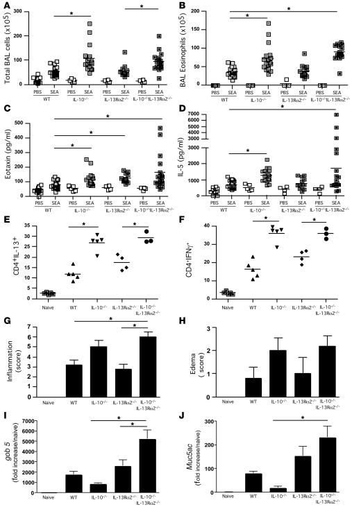
Figure 3. Exacerbated airway inflammation and airway pathology in IL-10–/–IL-13Rα2–/– dKO mice following SEA challenge.
Animals were sensitized and challenged as in Figure 1. Animals were euthanized 24 hours after the final airway challenge. *P < 0.05, 1-way ANOVA, except in Figure 3G, where a Kruskal-Wallis test was used. (A) Total airway infiltrates recovered in BAL. (B) Eosinophils recovered in BAL. (C) Eotaxin levels measured in BAL fluid by ELISA. (D) IL-5 levels measured by in BAL fluid by ELISA. (E) Lungs were excised, broken down into a single-cell suspension, and stimulated with PMA and ionomycin, with brefeldin A, for 3 hours and then stained with anti-CD4 (APC) and anti–IL-13 (PE) before cells were acquired and analyzed by FACS. (F) The procedure in F was identical to E apart from the final staining step using anti–IFN-γ and not anti–IL-13. (G) Perivascular/bronchial inflammation. H&E-stained sections from fixed and sectioned lungs were scored in a blinded fashion for cellular tissue infiltration. (H) Perivascular/bronchial edema. H&E-stained sections were scored in a blinded fashion for signs of edema. (I) RNA was extracted from lung tissue, with gob 5 mRNA quantified by qRT-PCR. (J) As in I, except Muc5ac mRNA was quantified.

