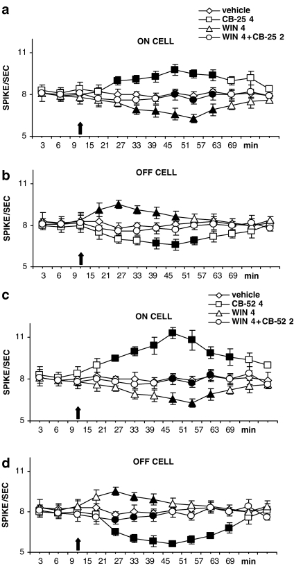
Figure 6.
Spontaneous activities of RVM ‘ON' (a, c) and ‘OFF' (b, d) cells before and after microinjections into the ventrolateral PAG of CB-25 (4 nmol per rat), CB-52 (4 nmol per rat) or WIN55,212-2 (4 nmol per rat) alone, or of CB-25 or CB-52 (2 nmol per rat) in combination with WIN55,212-2 (4 nmol per rat). CB-25 and CB-52 at the lowest dose (2 nmol per rat) did not change pro-nociceptive ‘ON' and anti-nociceptive ‘OFF' cell firing (for clarity, these curves are not shown). CB-25 and CB-52 at the highest dose increased pro-nociceptive ON cell firing (a, c) and decreased anti-nociceptive OFF cell firing (b, d). WIN55,212-2 (4 nmol per rat) caused effects that were opposite to those induced by CB-25 and CB-52 and were blocked by co-injection with the low, inactive, doses of CB-25 and CB-52 (2 nmol per rat). The black arrow denotes the time of injections of drugs. Each point represents the mean±s.e.m. of 10 recorded neurons. Significant differences between groups are shown as filled symbols (P<0.05; Wilcoxon signed rank test). For treatments with a single compound, means of the treated groups were compared to those from the relevant vehicle. For combination treatments, means were compared to those from treatment with the corresponding single compounds.

