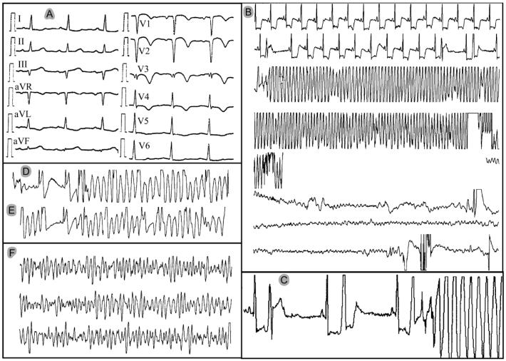
Figure 1.
Electrocardiograms of patient MMRL23 recorded during the first day of hospitalization. A: 12-lead electrocardiogram of evolving anterior myocardial infarction. B to F: single-lead monitor recordings. B: Episode of polymorphic VT deteriorating to VF. The transition from polymorphic VT to VF is missing because the monitor lead was disconnected during resuscitation. C: Onset of the VF episode shown in panel B. Note the accentuated ST depression and the short coupling interval (R-on-T phenomenon) of the ventricular ectopic beats, including the one initiating the polymorphic VT/VF. D and E: Additional episodes of non-sustained polymorphic VT starting with short-coupled extrasystoles. F: Additional VF episode that required electrical cardioversion (initiation not shown).

