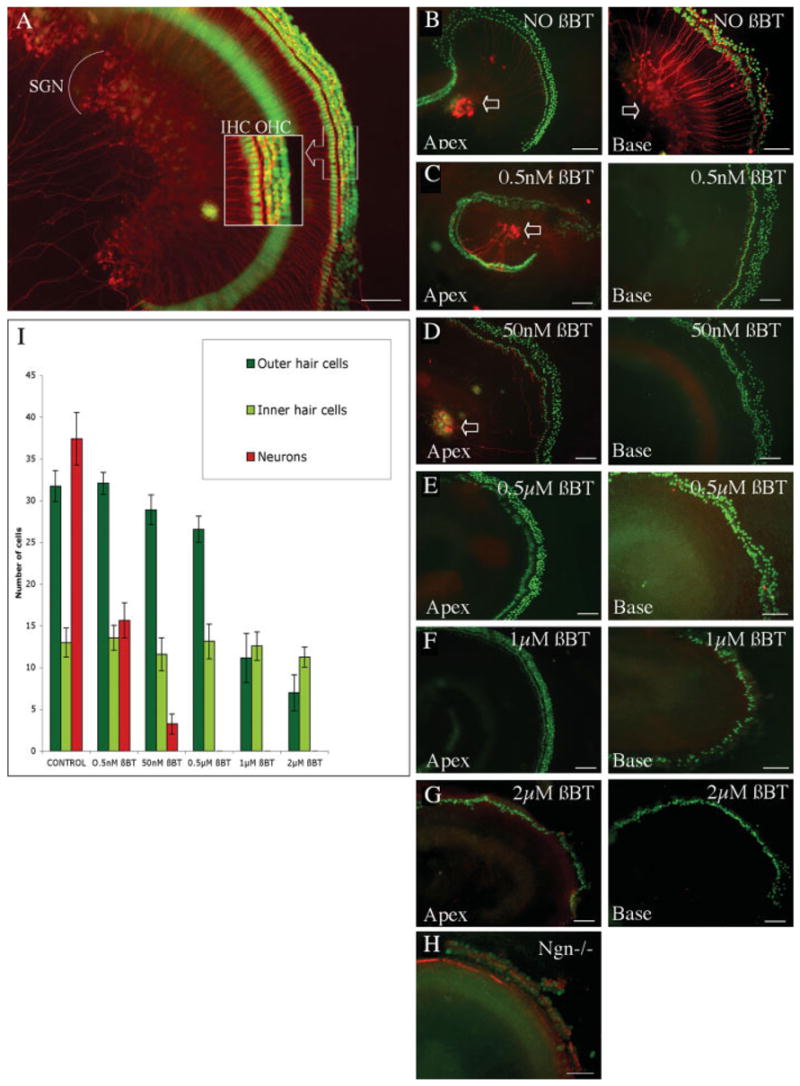
Figure 1.

Organ of Corti explants and treatment of explants with β-bungarotoxin. Organ of Corti explant of a C57BL/6 mouse after 3 days in culture (A). Outer hair cells (OHC) and inner hair cells (IHC) were visualized with an antibody to parvalbumin 3 and secondary FITC-labeled antibody (shown in green), and spiral ganglion neurons (SGNs) were detected with an antibody to β-III tubulin and TRITC-labeled secondary antibody (shown in red). Inset shows a higher magnification of the hair cells. After treatment for 2 days (B–G) in increasing concentrations of toxin (β-BT) organs of Corti of Atoh1-nGFP transgenic mice were immunostained with an antibody to β-III tubulin detected with a secondary TRITC-labeled antibody (shown in red) and hair cell nuclei were detectable by nuclear green fluorescence. The apical turns of the cochlea (Apex) are shown in the left column and basal turns (Base) are shown in the right column. (B) Untreated control explants showing intact hair cells and spiral ganglion neurons. Neurons stained for β-III tubulin (indicated by arrows). (C) Treatment with 0.5 nM β-bungarotoxin. Note that neurons at the base of the organ of Corti were eliminated but the neurons of the apex were not lost. (D) Treatment with 50 nM β-bungarotoxin. Note complete elimination of neurons at the base and partial survival in the apical turns. (E) Treatment with 0.5 μM β-bungarotoxin. There was no evidence of neuronal survival whereas the outer and inner hair cells were intact. (F) Treatment with 1 μM β-bungarotoxin. At this concentration there was significant loss of outer hair cells in the base of the cochlea and complete elimination of neurons. (G) Treatment with 2 μM β-bungarotoxin. Note that outer hair cells were nearly completely eliminated but inner hair cells were retained. Neurons were completely absent. (H) Organ of Corti explant of a newborn ngn 1 knock-out mouse. Immunostaining as above for neurons and with parvalbumin 3 using a FITC-labeled secondary antibody (shown in green) and phalloidin labeled with TRITC (shown in red) for hair cells. There was a complete lack of spiral ganglion neurons and a reduced number of hair cells. Scale bars in (A–H) are 100 μm. (I) Quantification of surviving outer hair cells, inner hair cells, and neurons from organ of Corti of Atoh1-nGFP transgenic mouse in culture for 48 h with different concentrations of β-bungarotoxin. Data are expressed as the mean number of cells ± SD per 100 μm of explant length. Higher concentrations of the neurotoxin decreased the number of outer hair cells. Beta-bungarotoxin (0.5 μM) for 2 days eliminated the neurons without significant alteration in the number of hair cells. [Color figure can be viewed in the online issue, which is available at http://www.interscience.wiley.com.]
