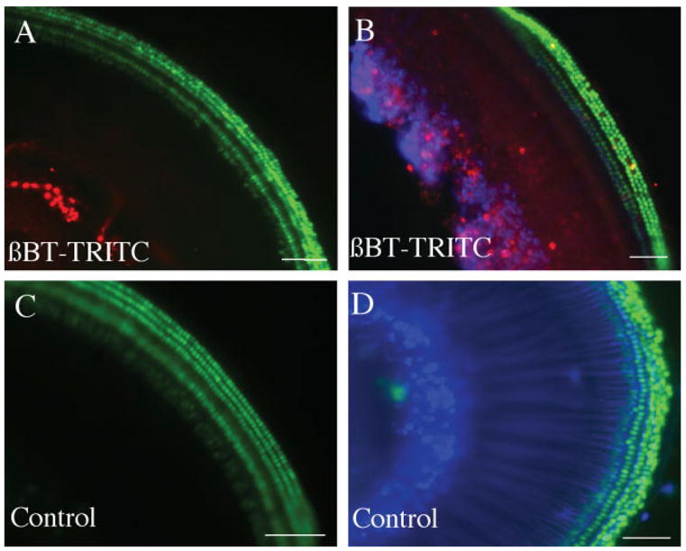
Figure 2.

Treatment of newborn mouse organ of Corti with TRITC-labeled β-bungarotoxin. Organs of Corti of Atoh1-nGFP transgenic mouse were treated with 50 nM β-bungarotoxin labeled with TRITC (shown in red) for 30 min (A,B). The controls were treated with medium without β-bungarotoxin but containing TRITC (C,D). Tissue was fixed without further staining (A,C) or immunostained with antibody to β-III tubulin followed by a Cy5-labeled secondary antibody [blue, (B,D)]. Note labeling by β-bungarotoxin-TRITC in the spiral ganglion neurons. Scale bars are 100 μm. [Color figure can be viewed in the online issue, which is available at http://www.interscience.wiley.com.]
