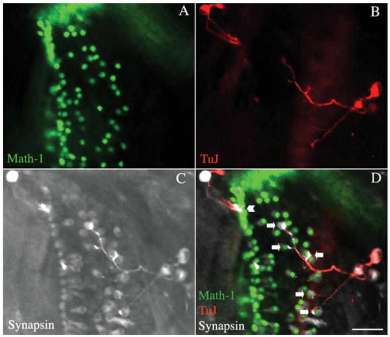
Figure 5.

Assessment of synaptic markers at points of contact between spiral ganglion neurons and hair cells in a recipient organ of Corti. The organ of Corti of an Atoh1-nGFP transgenic mouse was treated with β-bungarotoxin (A), and after 2 days, spiral ganglion neurons obtained at P1 by dissociation of the tissue from a C57BL/6 mouse were added to the organ of Corti for 2 days. Hair cells visualized by endogenous fluorescence (green). (B) Staining with an antibody to β-III tubulin followed by a TRITC-labeled secondary antibody (shown in red). (C) Staining with antibody to synapsin detected with a Cy5-labeled secondary antibody (shown in white). (D) Merged image. The contacts between transplanted neurons and hair cells were immunopositive for synapsin. Neurons made several contacts with outer hair cells (arrows), while they made single contacts with inner hair cells (arrowhead). Scale bar is 30 μm. [Color figure can be viewed in the online issue, which is available at http://www.interscience.wiley.com.]
