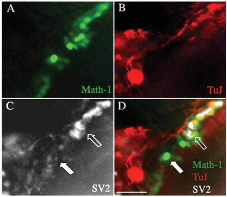
Figure 6.

Staining of synaptic markers in neurons cocultured with a β-bungarotoxin treated organ of Corti. An organ of Corti from an Atoh1-nGFP transgenic mouse was treated with β-bungarotoxin (A) and dissociated spiral ganglion tissue from a P1 mouse was added for a 2-day culture. Hair cells were positive for endogenous GFP. (B) Staining with an antibody to β-III tubulin followed by a TRITC-labeled secondary antibody (shown in red). (C) SV2 antibody detected with a Cy5-labeled secondary antibody (shown in white). (D) Merged image. Strong staining for SV2 was seen at points of contact between neurons from the C57BL/6 mouse and hair cells from the Atoh1-nGFP mouse (open arrow). No staining was seen in hair cells that were not in contact with a neuron (solid arrow). Scale bar is 30 μm. [Color figure can be viewed in the online issue, which is available at http://www.interscience.wiley.com.]
