
FIGURE 2.
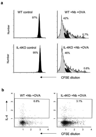
In vivo Ag-specific T cell expansion and cytoplasmic IL-4 levels are pronounced in the absence of non-T cell or bystander T cell IL-4. Five million purified DO11.10 CD4+ T cells were labeled with CFSE and transferred to recipient BALB/c IL-4–/– mice or WT mice. Two days later, recipient mice were immunized intracutaneously in the ear with N. brasiliensis (Nb) and OVA peptide (five animals per treatment group). At day 7 after immunization, the draining CLN cells were collected. a, Analysis of cell cycling was performed as described in Fig. 1. b, For ex vivo cytoplasmic cytokine staining, CLN cells were cultured with 10 μg/ml OVA peptide for 6 h with GolgiStop added for the last 4 h. Intracellular staining of IL-4 was performed as described in Materials and Methods. Data shown are for gated CD4+KJ1-26+ OVA-specific T cells. This experiment was repeated three times with similar results.