
FIGURE 3.
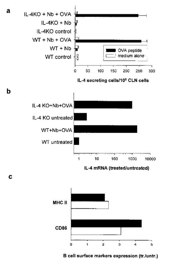
Transferred Ag-specific T cells effectively differentiated into IL-4-secreting cells in IL-4–/– mice. The DO11.10 T cell transfer and inoculation of recipient mice was performed as described in Fig. 2. a, CLN cells were cultured for 3 days with or without OVA restimulation to assess OVA-specific IL-4 secretion by ELISPOT assay. b, Transferred OVA-specific DO11.10 T cells were purified from CLN cells by incubating with KJ1-26-PE and anti-PE beads as described in Materials and Methods. IL-4 mRNA expression in sorted DO11.10 T cell populations from pooled treatment groups (five animals per group) was determined by quantitative TaqMan real-time PCR. c, CLN cells were pooled (five mice per treatment group) and stained with anti-B220 and anti-MHC II or anti-CD86 Abs. Expression of MHCII (mean fluorescence intensity) and CD86 (percentage of positive B cells) on gated B220+ cells were assessed by FACS analysis. Treatment groups included: IL-4–/– recipient mice (■) or WT recipient mice (□) inoculated with N. brasiliensis (Nb) + OVA, expressed relative to untreated recipient IL-4–/– mice or untreated recipient WT mice, respectively. This experiment was repeated twice with similar results.