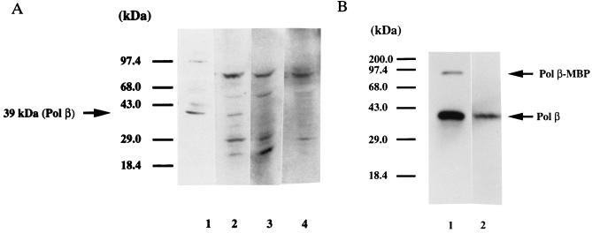
Figure 1.
The antiserum raised against a pol β–MBP fusion protein specifically recognizes pol β. (A) Western blot. Lane 1, 100 ng of a partially purified fraction (from E. coli) of recombinant rat DNA pol β detected with pol β antiserum at 1:300 (larger band is residual uncleaved pol β-MBP). Lanes 2–4, whole testicular nuclei (6 μg). Lane 2, pol β antiserum at 1:300; lane 3, pol β-depleted serum at 1:50; lane 4, preimmune serum at 1:50. (B) Activity gel analysis. Lane 1, partially purified recombinant rat DNA pol β (100 ng) from E. coli; lane 2, testicular nuclei (50 μg).

