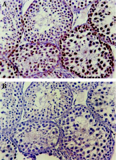
Figure 2.
Differentiating mouse spermatocytes stain with antiserum to pol β. (A) Immunohistochemical staining of seminiferous tubule of a normal 3-week-old mouse labeled with pol β IgG (60 μg/ml). Mouse spermatocytes show significant brown staining with the pol β IgG, while the interstitial cells and Sertoli cells do not stain brown. (B) Immunohistochemical staining of seminiferous tubule from a normal 3-week-old mouse incubated with pol β-depleted IgG (60 μg/ml). No brown staining was detected.

