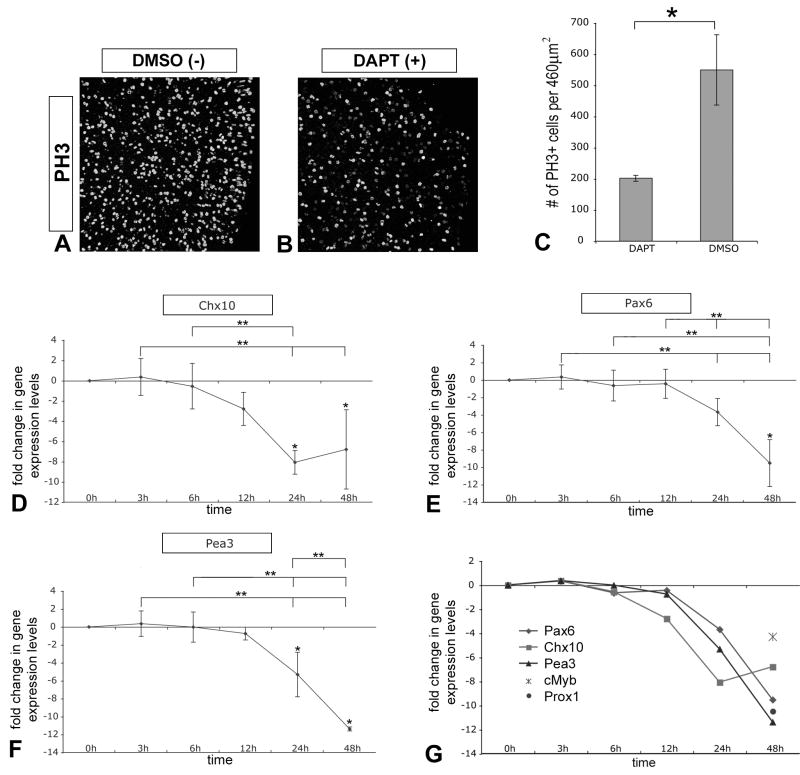
Figure 2. Loss of Notch signaling reduces proliferation and progenitor gene expression.
(A, B) E4.5 chick retinal explants pairs cultured for 48h in DAPT or DMSO were wholemount immunolabeled with anti-phospho Histone 3 (PH3) antibody to reveal mitotic progenitor cells at the apical surface of the retina: images were acquired from flatmounted explants with LSCM. (C) Quantification of PH3+ progenitor cells indicated that DAPT treatment significantly reduced proliferation ∼3-fold compared to control; error bars indicate standard error of the mean, n=3 pairs, P<0.0186. (D–F) QPCR was used to analyze changes in Chx10 (D), Pax6 (E), and Pea3 (F) gene expression levels over time due to DAPT treatment as described before. (G) Relative comparison of changes in progenitor gene expression levels over time, including Prox1 and cMyb levels at the 48h timepoint. Note that decline in expression levels are apparent by 24h of Notch signaling inactivation.

