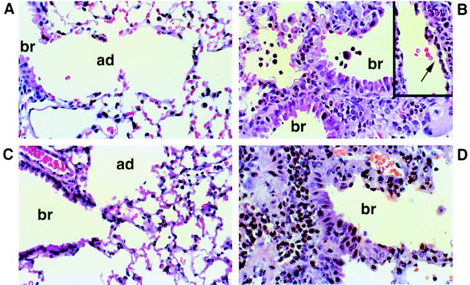
Figure 3.
Lung histology from wild-type (WT) and IgE-deficient mice (KO) after treatment with NS or Af. (×600.) (A) WT mouse treated with NS. (B) WT mouse treated with intranasal Af extract shows a peribronchiolar inflammatory infiltrate consisting predominantly of eosinophils, admixed with lymphocytes. Involvement of the bronchiolar epithelium with associated epithelial damage is present. The airspaces contain numerous eosinophils and histiocytes. Vessels are surrounded by a cuff of inflammatory cells and contain marginating eosinophils with migration into the vessel wall (arrow in Inset). (C) KO mouse treated with NS. (D) KO mouse treated with intranasal Af shows essentially the same findings as those seen in WT mice. Br, bronchiole; ad, alveolar duct. These panels depict fields representative of the histology obtained from six or seven mice per group.

