Figure 1.
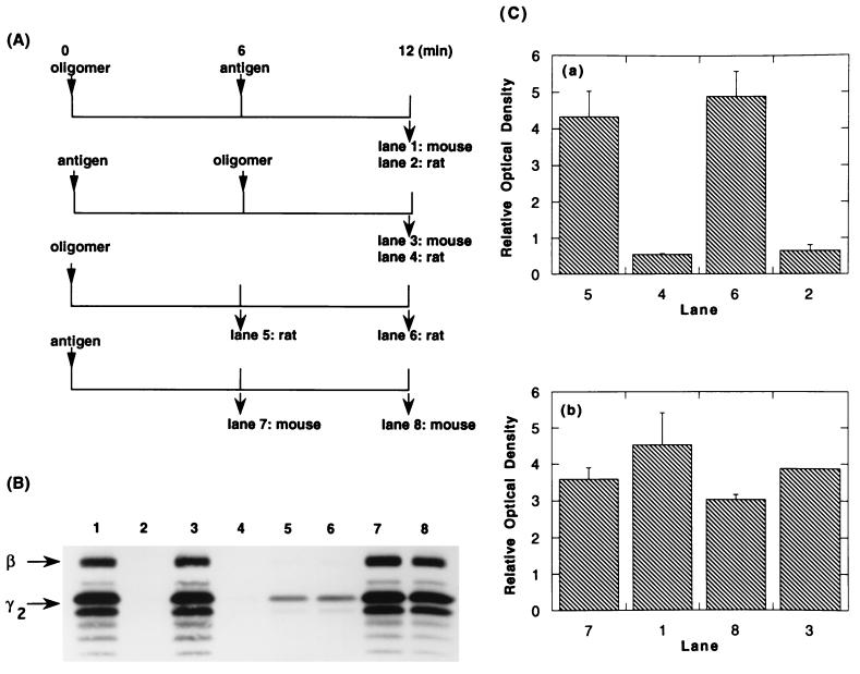
Effect of aggregating IgE-FcɛRI with antigen on phosphorylation of receptors previously aggregated by IgE oligomer. (A) Protocols for stimulating cells and immunoprecipitating their receptors. FcɛRI on RBL-2H3 cells were partially (52%) saturated with DNP-specific monomeric mouse IgE and reacted at 6.25 × 10⁶ cells/ml with rat IgE oligomer (0.3 μg/ml) or antigen (DNP6-BSA, 100 ng/ml) or both. At each time point, the total IgE was immunoprecipitated with species-specific anti-IgE, and the phosphotyrosine on the subunits of the receptor was analyzed. (B) Autophotograph of a Western blot using antiphosphotyrosine antibodies. Each lane contains approximately 5 × 10⁶ cell equivalents. (C) Densitomeric quantitation of the blot shown in B. The bars show the averages of two samples, and the ranges of the duplicates are shown. The ordinate values are graphed on a relative scale; the absolute ratio of the data for b:a was as 15:1. In numerous experiments of this type the ratio approximated 10:1.

