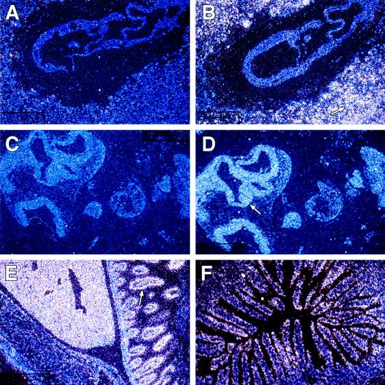
Figure 4.
The temporal and spatial pattern of Ell expression during murine development. In situ hybridization studies were performed with radiolabeled Ell 3′ UTR sense (A and C) and antisense (B, D, E, and F) riboprobes. (A and B) Hybridization to longitudinal sections of E7.5 embryonic tissues (×10). The arrow indicates the maternally derived decidual tissue that exhibited high levels of Ell expression. (C and D) Hybridization to longitudinal sections of E9.5 embryonic tissues (×10). Although expression is diffuse throughout the embryo at E9.5, it is slightly more prominent at the neuroepithelium as indicated by the arrow. (E) Hybridization to a longitudinal section of the abdominal cavity of a 1-day-old mouse (×1.25). The abdominal wall is at the lower left and exhibits only minimal Ell expression. In the middle of the panel, a high level of expression is seen in the liver. The arrow indicates expression in the intestine. (F) Hybridization to a cross-section of adult intestine (×10) and illustrates the localization of Ell expression to the intestinal villi.

