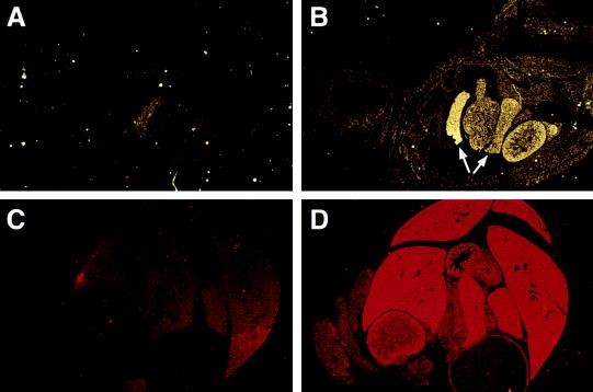
Figure 5.
In situ hybridization studies were performed with radiolabeled Ell 3′ UTR sense (A and C) and antisense (B and D) riboprobes. (A and B) Hybridization to longitudinal sections of E16.5 embryonic tissues (×1.25). The arrows indicate expression in lobes of the liver. The gastrointestinal tissues, which are adjacent to the liver, also display prominent expression. (C and D) Hybridization to cross-sections of the abdominal cavity of a 1-day-old mouse (×1.25). Prominent expression can be appreciated in the hepatic and gastrointestinal tissues with the antisense probe compared with the sense control.

