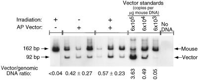
Figure 4.
Quantitative PCR of vector DNA in mouse liver DNA. Amplification of vector DNA and mouse genomic DNA was performed to quantify the relative levels of AP vector DNA for the following samples. Mice were treated with 1 × 10⁵ ffu of CWRAPSP, either with or without previous exposure to 17 Gy of γ-irradiation. Liver DNA for a mouse injected with AAV-LIXSN and exposed to 17 Gy served as a negative control for vector DNA amplification. The indicated amounts of virion DNA were added to 1 μg of the negative-control sample to generate standards for vector quantitation. No DNA was added to the last sample. Signals were quantified with a phosphorimager. The signal for the vector DNA (92 bp) was normalized to the signal for mouse genomic DNA (162 bp) to correct for varying efficiencies of amplification. The ratio of vector (cDNA) signal to genomic signal and the SD for each condition are shown.

