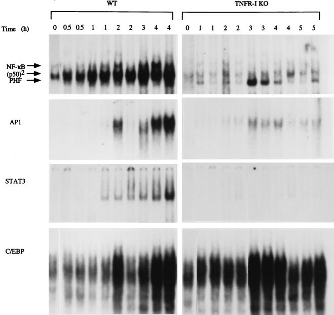
Figure 1.
Transcription factor binding after PH in wild-type (WT) and TNFR-I knockout mice (TNFR-I KO). Mice were partially hepatectomized and killed 0.5 and 5 h after the operation, as indicated at the top of the figure (two mice per time point). Nuclear extracts were prepared, and EMSAs were performed using 10 μg of nuclear protein in each lane. Reticulocyte lysate was used as a marker to determine the position of the p50/p65 NF-κB heterodimer (11). p50 homodimer position is indicated as (p50)2. There was variability in AP-1 binding in wild-type mice killed 2 h after PH. PHF, PH factor

