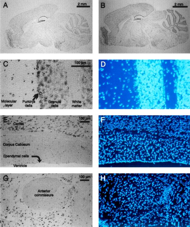
Figure 4.
In situ hybridization of mBNaC1 (A, E, and G) and mBNaC2 (B and C) riboprobes to mouse brain sagittal sections, and DAPI stain of nuclei in those same sections (D, F, and H). Glial cell nuclei are brighter. (A and B) Whole brain. (C and D) Portion of cerebellum with white matter, granule cell layer, Purkinje cell layer, and molecular layer indicated. (E and F) Portion of parietal cortex, corpus callosum, ependymal cell layer, and ventricular space. (G and H) Anterior commissure and surrounding neuronal areas. Hybridization of control sense riboprobes to adjacent sections under the same conditions gives no signal (not shown). In all pictures (except for C and D, whose orientation has not been determined), anterior is to the left and ventral is down.

