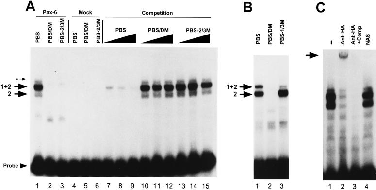
Figure 2.
Binding of Pax-6HA protein to the N-CAM PBS. (A) Pax-6HA expressed in COS-1 cells (lanes 1–3) and mock-transfected COS-1 cell extracts (lanes 4–6) were incubated with either 32P-labeled PBS (lanes 1, 4, and 7–15), PBS/DM (lanes 2 and 5), or PBS-2/3M (lanes 3 and 6). Complexes formed by Pax-6HA and the PBS probe are indicated by arrows labeled 2 and 1+2. A minor complex formed by both the Pax-6HA and mock-transfected COS-1 extracts is indicated by an asterisk. A 100-, 250-, or 500-fold molar excess of either unlabeled PBS (lanes 7–9), PBS/DM (lanes 10–12), or PBS-2/3M (lanes 13–15) were added as competitors. (B) Pax-6HA extracts were incubated with the PBS probe (lane 1), PBS/DM (lane 2), or PBS-1/3M (lanes 3). DNA/protein complexes are indicated by arrows labeled 2 and 1+2. (C) Supershift analysis. Pax-6HA was incubated with the 32P-labeled PBS probe and either 1 μl of HA tag antibody (lanes 2 and 3), an antibody to Ng-CAM (NAS) (lane 4), or no antibody (lane 1). In lane 3, 500-fold excess of unlabeled PBS was added as competitor. The supershifted Pax-6/PBS complex is indicated by the arrow.

