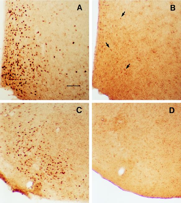
Figure 4.
Photomicrographs showing the ER–IR cells (stained with polyclonal ER21 antibody) in the medial preoptic area and the ventromedial nucleus of hypothalamus of WT (A and C) and ERKO (B and D) mouse brains. The number of ER–IR cells was greatly reduced in ERKO mouse brains but not completely eliminated in the medial preoptic area as indicated with arrows in B. (Scale bar = 75 μm.) Despite the evident loss of ER–IR cells, there was no overall difference on the distribution of AR–IR cells between ERKO and WT mice (see Table 1).

