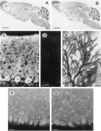
Figure 2.
Histological analysis. Midsagittal section of wild-type (A) and calbindin null mutant (B) mice stained with cresyl violet. Purkinje cells of wild-type (C) and null mutant (D) mice stained with antiserum to calbindin. (E) Golgi staining of Purkinje cell dendrites shows normal spine morphology and density. Purkinje cells of wild-type (F) and mutant (G) mice visualized with antiserum to parvalbumin.

