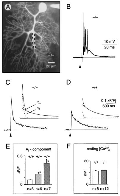
Figure 4.
Synaptically evoked calcium transients in cerebellar Purkinje neurons from mice lacking calbindin. (A) Confocal fluorescence image of a Purkinje neuron in a cerebellar slice obtained from a 24-day-old null mutant mouse (−/−). The neuron was loaded through the recording patch pipette with the calcium indicator dye, Calcium Green-1. The region delimited by the dashed line indicates a typical dendritic area from which the fluorescence data (C and D) were obtained. (B) Excitatory postsynaptic potential (complex spike) evoked by extracellular stimulation of the afferent climbing fiber. Recording from a null mutant mouse, membrane potential (Vm) was −70 mV. (C and D) Dendritic calcium transients evoked by climbing fiber stimulation recorded in null mutant mice (C, −/−, average trace from n = 7 cells in 4 mice) and wild-type mice (D, +/+, average trace from n = 6 cells in 4 animals). The curves above each fluorescence trace represent superpositions of the corresponding double-exponential time-course fit (continuous line) with the fast (tf, dotted line) and the slow (ts, dashed line) components depicted separately. (E) Af amplitude components (corresponding to tf) of the synaptically induced calcium signals in wild-type, heterozygous (+/−) and null mutant mice, respectively. (F) Free intracellular calcium concentration ([Ca2+]i) under resting conditions measured in somata of fura-2 loaded cells (50 μM) of wild-type and null mutant mice. In E and F, values are the means ± SEM. The asterisk indicates significant difference to wild type (Student’s t test, P < 0.05). Arrowheads mark the time points of single-shock synaptic stimulation.

