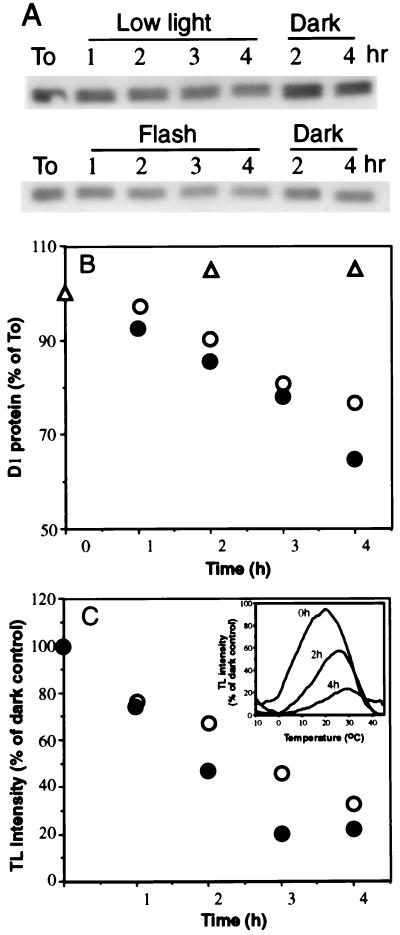
Figure 3.
Kinetics of the PSII inactivation and D1 protein degradation induced by single turnover flashes. Thylakoids were exposed to flashes delivered at 40-s dark interval (•), to continuous white light (○, 30 μmol m−2·s−1) or as a control were kept in the dark for the same period (▵). Lane To, D1 protein level before light exposure. All incubations were at room temperature. Sample where taken at times as indicated and assayed for the D1 protein content (A and B) and TL signal (C). (C Inset) Recorded TL measurements indicating loss of the QB•−/S2,3 emission (22) as a function of increasing exposure time to the flashes. No block in electron flow from QA•− to QB or loss of the oxygen evolving complex activity occurred as evidenced by the absence of the QA•−/S2 band (emission at 7–10°C).

