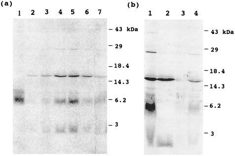
Figure 3.
Labeling of pea mitochondrial proteins with [2-14C]malonic acid. Pea mitochondria were labeled with [2-14C]malonic acid, and proteins were separated by SDS/PAGE, blotted onto nitrocellulose membrane, and analyzed by PhosphorImager. (a) Time course of labeling of mitochondrial proteins and effect of ATP and MgCl2. In lanes 2–5, mitochondria were labeled for 10, 20, 40, and 60 min, respectively. In lanes 6 and 7, mitochondria were labeled for 1 h, but ATP or MgCl2, respectively, were omitted from assay medium. Lane 1 shows a standard of 14C-labeled 16:0-mtACP. (b) Effects of DTT treatment on deacylation of labeled proteins and cerulenin on labeling of proteins. Pea mitochondria were labeled for 1 h. Lane 1, control that was not treated with DTT; lane 2, mitochondrial proteins were treated with DTT; lanes 3 and 4, 200 μM and 20 μM cerulenin, respectively, were added to the incubation medium, and samples were not treated with DTT.

