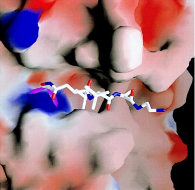
Figure 5.
Close-up view of the active-site cleft of TACE. On top of the solid surface representing the proteinase the bound inhibitor is shown in full structure, slotting with its isobutyl (P1′) and its Ala (P3′) side chains into the deep S1′ and the novel S3′ pockets. Figure was made as Fig. 2b (22).

