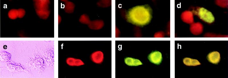
Figure 3.
Subcellular distribution and colocalization of hVIP/MOV34 and Vpr by indirect immunofluorescence assay. (a–d) HeLa cells were transfected with either hVIP/MOV34 or HIV-1 Vpr expression vectors, fixed for immunofluorescence as described previously (28), probed with anti-Vpr (a and c) and anti-Xpress antibody (b and d), and stained with fluorescein-conjugated goat anti-rabbit for Vpr or goat anti-mouse secondary antibody for hVIP. hVIP localized to the nucleus with a punctate staining pattern and Vpr localized to the periphery of the nucleus. a and b were vector-transfected; c, Vpr-transfected; d, hVIP/MOV34-transfected. (e–h) Colocalization of hVIP/MOV34 and Vpr. HeLa cells were cotransfected with hVIP/MOV34 and HIV-1 Vpr expression vectors and then fixed for immunofluorescence. Fixed cells were probed with anti-rabbit polyclonal primary antibody followed by staining with rhodamine-conjugated goat anti-rabbit secondary antibody for Vpr. The cells were probed with anti-Xpress mAb, followed by fluorescein-conjugated goat anti-mouse secondary antibody for hVIP/MOV34. (e) Phase-contrast field. (f) Rhodamine-specific Vpr fluorescence. (g) Fluorescein-specific hVIP/MOV34 fluorescence. (h) Double exposure in which the mixture of rhodamine and fluorescein appears yellow.

