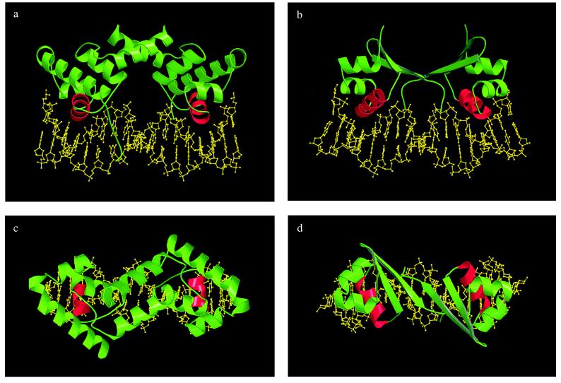
Figure 1.
Comparison of the structures of complexes of λ-repressor (5, 6) and Cro (unpublished results) with operator DNA. In both cases the proteins were crystallized with a 19-bp duplex of which the central 17 bps are shown. Recognition helices are shown in red. (a) Headpiece of λ-repressor bound to DNA. The consensus half is to the left. As can be seen there is substantial asymmetry, especially in the location of the amino-terminal “arm.” (b) Cro bound to operator DNA. (c) View down the pseudo 2-fold axis of λ-repressor bound to operator DNA. (d) Related view of Cro bound to operator DNA.

