Figure 2.
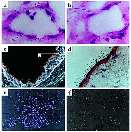
CXCR3 mRNA expression by endothelial cells from kidney of patients suffering from glomerulonephritis. (a) CXCR3 mRNA expression in the kidney biopsy specimen. The section, which was hybridized with 35S-labeled CXCR3 antisense probe, shows positive signal in a vessel wall. ×1,000. (b) Autoradiograph of a consecutive section hybridized with sense CXCR3 probe, showing virtually no signal. ×1,000. (c) Association between CXCR3 mRNA expression and vWF, as shown by combining in situ hybridization with CXCR3 antisense probe (white grains along the vessel wall) and immunohistochemistry with anti-vWF mAb (red) (dark field, ×250). (d) Higher-power magnification (×1,000) of inset in (c) showing both CXCR3 mRNA (black grains) and vWF expression (red). (e) CXCR3 mRNA expression by Th1 cells, used as positive control. The cells were hybridized with antisense CXCR3 probe (dark field, ×250). (f) Autoradiograph of the same Th1 cell culture hybridized with sense CXCR3 probe, showing virtually no signal (dark field, ×250).

