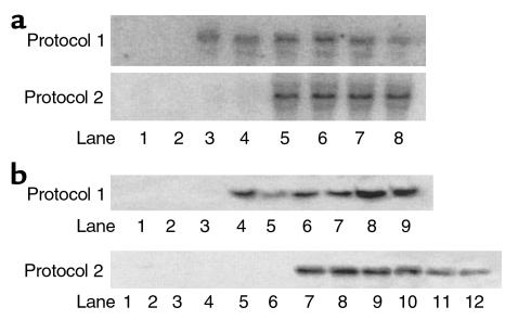
Figure 3.
Northern and Western blot analyses of 6PF-2K/F-2,6-P2ase overexpression. (a) Northern blot analysis of 6PF-2K/F-2,6-P2ase mRNA. Total RNA was extracted from fresh liver of all mice on day 7 after adenovirus infusion. For both protocols, only three animals of each group were chosen, without bias, and used for Northern blot analyses. A total of 30 μg RNA was loaded in each lane for electrophoresis. In protocol 1, samples were from normal mice treated with adenovirus (two representative animals of Ad-gal group, lanes 1 and 2; three animals of Ad-Bif-WT group, lanes 3–5; and three animals of Ad-Bif-DM group, lanes 6–8). In protocol 2, samples were from saline control mice and STZ-induced diabetic mice treated with adenovirus (two representative animals of each group are shown in blots). Saline control group, lanes 1 and 2; STZ-Ad-gal group, lanes 3 and 4; STZ-Ad-Bif-WT group, lanes 5 and 6; and STZ-Ad-Bif-DM group, lanes 7 and 8). (b) Western blot analysis of 6PF-2K/F-2,6-P2ase protein. Extracted protein was prepared from fresh liver tissue of mice sacrificed on day 7 after adenovirus infusion. For both protocols, only three animals of each group were chosen, without bias, and used for Western blot analyses. Concentration of extracted protein was measured by the BCA method. Equal amounts of extracted protein (100 μg) were used for electrophoresis. In protocol 1, samples were from the Ad-gal group (lanes 1–3), Ad-Bif-WT group (lanes 4–6), and Ad-Bif-DM group (lanes 7–9). In protocol 2, samples were from the saline control group (lanes 1–3), STZ-Ad-gal group (lanes 4–6), STZ-Ad-Bif-WT group (lanes 7–9), and STZ-Ad-Bif-DM group (lanes 10–12).

