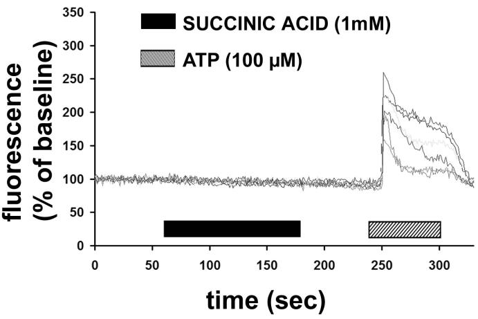
Figure 5.
Succinate does not increase cytosolic Ca2+ in hepatic stellate cells. Graphic shows representative tracings of 7 separate quiescent stellate cells serially stimulated with succinate (1 mM), and then the positive control ATP (100 μM). Cells were loaded with the fluorescent Ca2+ dye Fluo-4/AM, and then monitored using time-lapse confocal microscopy. Increases in free cytosolic Ca2+ were measured and are represented as increases in fluorescence intensity relative to baseline. Results are representative of what was observed in 132 separate stellate cells from 3 separate preparations.

