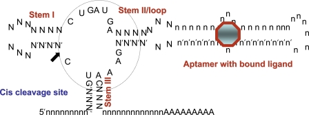
Fig. 1.
The cis-cleaving hammerhead ribozyme with aptamer and ligand. The three helical stems of the ribozyme are indicated, as is the catalytic core of the ribozyme and the site of cis cleavage. The position of an aptamer is indicated. When the ligand (octagon) is bound to the aptamer, it propagates a structural change in the catalytic center of the ribozyme, allowing it to cleave in cis. The catalytic core of the ribozyme is circled.

