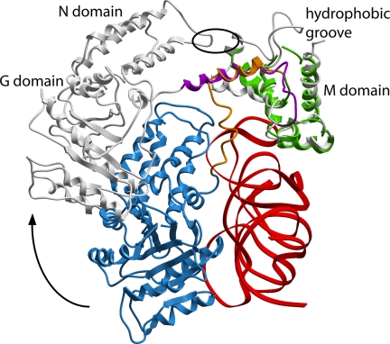
Fig. 5.
Superposition of the SRP54 M domains of the S. solfataricus SRP54–helix 8 complex (gray) and the M. jannaschii S domain A complex. Shown is the shift in position of the NG domain and the formation of the long GM-linker helix (magenta) when the NG domain is released from the RNA. The color code in the M. jannaschii S domain is as in Fig. 1. SRP19 is omitted for clarity. The position of the hydrophobic contact between the apical loop in the N domain and the finger loop in the M domain in the S. solfataricus SRP54–helix 8 structure is indicated by an oval.

