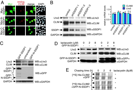
Fig. 2.
A cascade of protein interactions protects LIM-HD complexes from proteasomal degradation. (A) αT3 cells were transfected with Myc-SSDP1 expression constructs. Cells are costained with Myc (red) and CLIM (green) antibodies. Note the higher levels of endogenous CLIM in nuclei of cells transfected with Myc-SSDP1 and Myc-N-SSDP1 (1–92) containing the CLIM-interaction domain, but not Myc-C-SSDP1 (90–361). (B) Knock-down of SSDP1 results in decreased levels of endogenous CLIM and Lhx3. SSDP1 levels were knocked-down in αT3 cells via retroviral infection of three independent mouse shRNAs directed against different regions on SSDP1 or the empty retroviral vector as control. Note that knocking down endogenous SSDP1 levels in αT3 cells leads to a decrease in endogenous CLIM and Lhx3 protein levels (Left), whereas mRNA levels remain unchanged as measured by qRT-PCR (Right), (n = 3; values are mean ± SE). (C) Western blot of protein extracts from αT3 cells transfected with GFP-SSDP1 and GFP-N-SSDP1 expression constructs. GFP-positive cells were isolated via FACS before extract preparation. GFP-negative cells from the same sorting were used as negative control. The same blot was probed with antisera against Lhx3, CLIM, GFP, and GAPDH. Note the higher levels of endogenous CLIM and Lhx3 in cells overexpressing GFP-SSDP1 or GFP-N-SSDP1. (D) SSDP1 protects the Lhx3-CLIM complex from proteasomal degradation. αT3 cells were transfected with GFP-N-SSDP1 expression construct, and GFP-positive cells were isolated via FACS. Both GFP-positive and GFP-negative cells of the same sorting were cultured overnight. Cells were then treated with indicated concentrations of lactacystin for 6 h before harvesting for Western blotting. The same blot was probed with antisera against Lhx3, CLIM, and GFP/GAPDH. Note that Lhx3 and CLIM protein levels in GFP-N-SSDP1-expressing cells are no longer sensitive to lactacystin. (E) SSDP1 increases the half-life of CLIM. His-tagged CLIM2 was coexpressed in HEK293T cells with GFP or GFP-N-SSDP1 and total cellular proteins were labeled with [35S]methionine for 1 h, followed by washings and chasing in normal medium for indicated time periods in the presence or absence of lactacystin.

