Fig. 3.
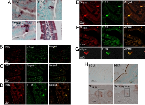
Detection and localization of T1R receptors and Gαgust along the crypt–villus axis of small intestine. (A) In situ hybridization with antisense riboprobes to T1R2, T1R3, and Gαgust. Complementary sense probes to all targets did not hybridize to any transcripts within the tissue sections (see SI Fig. 6). (B and C) Immunofluorescent detection of the T1R3 taste receptor subunit (red) and Gαgust (green) in serial wax sections of mouse duodenum. (D) Immunofluorescent detection of the T1R3 (red) and T1R2 (green) taste receptor subunits in a single wax section of human duodenum. (E–G) Immunofluorescent detection of the T1R2 and T1R3 taste receptor subunits (green), and Gαgust (red) in a single wax section of human duodenum. (H) Chromogenic detection of SGLT1 in wax sections of mouse small intestine. (I) Chromogenic detection of Gαgust and chromogranin in serial wax sections of mouse proximal intestine. The boxed cell expresses both Gαgust and chromogranin.

