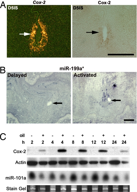
Fig. 4.
Uterine expression of miR-101a, miR-199a*, and Cox-2. (A) In situ hybridization of Cox-2 with 35S-labeled probe (Left) and protein by immunohistochemistry (Right) in serial cross-sections from day-5 implantation sites (IS). (B) In situ hybridization of miR-199a* with DIG-labeled LNA probe on longitudinal sections from delayed and implanting uteri. Arrows indicate the location of blastocysts. (C) Uterine Cox-2 and miR-101a levels during experimentally induced decidualization. Western blot shows Cox-2 protein levels at time points after intrauterine oil infusion (+); noninfused contralateral uterine horns serve as negative controls (−). Actin is a loading control. The ethidium bromide-stained gels served as loading control for miR-101a Northern blot. (Scale bars: 200 μm, A; 400 μm, B.)

