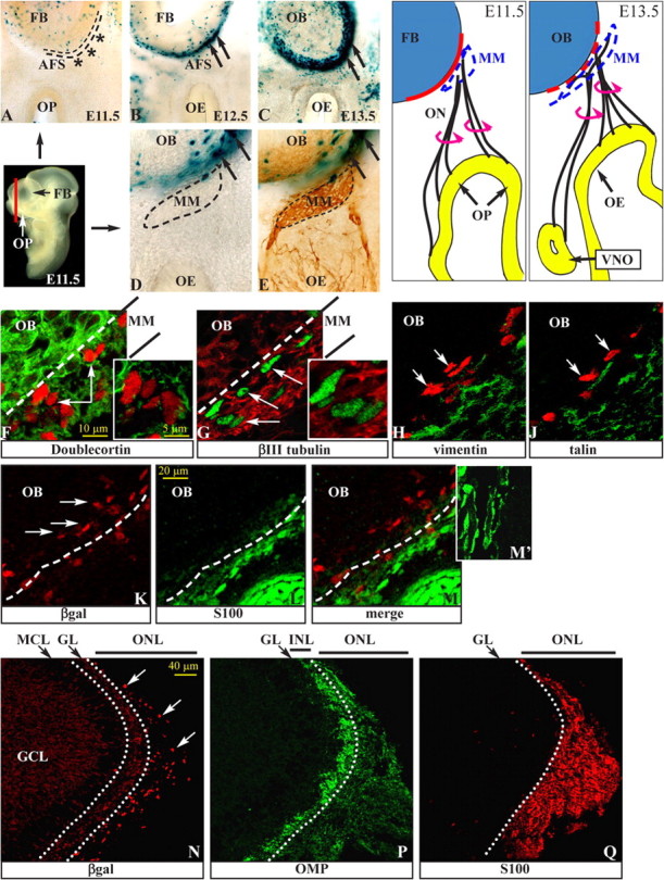
Figure 1.

Wnt–βcatenin-responsive cells in the developing olfactory system. A–C, X-gal staining of frontal sections of BATnlacZ embryos at E11.5 (A), E12.5 (B), and E13.5 (C). A section plane is shown (solid red line). βgal+ nuclei are visible around the OB at E12.5 and subsequent stages (black arrows) but not at E11.5 (black asterisks). D, E, X-gal staining of sections from E12.5 BATnlacZ embryos (D), followed by immunostaining with anti-NCAM (E), to show the position of the MM (area within the dashed line) and the βgal+ nuclei. The drawings on the right show the relationship between ORN neurites and the FB at the same stages. F–J, Double immunostaining for βgal (white arrows) and DCX (F, red), βIII-tubulin (G, green), vimentin (H, green), or talin (J, green) on sections of E12.5 BATnlacZ embryos. Insets, Higher magnification of single confocal Z-slices. K–M, Double immunostaining for βgal (K, M, red) and S100 (L, M, green). M′, Higher magnification of ON-associated S100+ cells. N–Q, Immunostaining for βgal (N, red, white arrows), OMP (P, green), and S100 (Q, red) on consecutive sagittal sections of neonatal BATnlacZ;Dlx5+/lacZ OB. In N, anti-βgal also recognizes OB interneurons (weaker cytoplasmic signal). Dotted white lines separate the outer and the inner nerve sublayers. GCL, Granule cell layer; GL, glomerular layer; INL, inner nerve layer; MCL, mitral cell layer; ONL, outer nerve layer.
