
Figure 2.
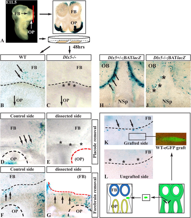
Organotypic cultures to define the origin of Wnt signal. A, Section plane and experimental procedure using organotypic cultures. B, C, Sections of E11.5 BATnlacZ WT (B) and BATnlacZ;Dlx5−/− (C) embryos, cultured for 48 h and stained with X-gal. βgal+ nuclei (black arrows) are visible in the WT but not in the Dlx5−/− (black asterisks). D, E, Removal of one OP (dashed red line) from slices of E11.5 BATnlacZ WT embryos: βgal+ nuclei are detected in the control sections (black arrows) but absent after OP removal (black asterisks). F, G, Same as in D and E but removing the FB (dashed red line). The number of βgal+ nuclei is not greatly reduced compared with the control side. H, J, Coronal sections of E13.5 embryos with genotypes BATnlacZ;Dlx5+/− (H) and BATnlacZ;Dlx5−/− (J), stained with X-gal. The Dlx5−/− sections show a drastic reduction in βgal+ nuclei (black asterisks) compared with the Dlx5 heterozygous ones (black arrows). K, L, Grafting of fragments of OP from E11.5 eGFP+ WT donor embryos onto slices from E11.5 BATnlacZ;Dlx5−/− embryos. The donor tissue was placed anterior to the FB, as illustrated in the drawing (bottom) and indicated in K. After culture, βgal+ nuclei were detected (X-gal staining) in the AFS of grafted slices (arrows in K) but not in the control side (asterisks in L). NSp, Nasal septum.