
Fig. (6). Nuclear DNA fragmentation occurs in conjunction with phosphorylation of the retinoblastoma protein (Rb) following NO exposure.
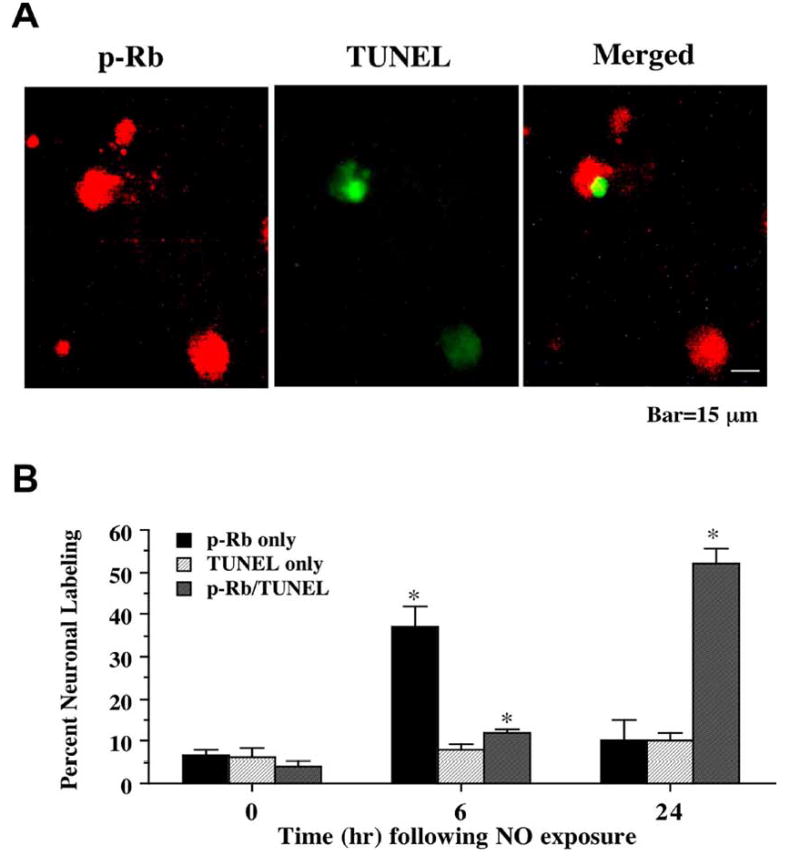
(A) Representative fields illustrate the double staining of neurons with p-Rb and TUNEL performed 24 hr following NO exposure (NOC-9, 300 μM). NO exposure resulted in significant p-Rb staining (red). p-Rb labeling (green) also was evident in neuronal cultures exposed to NO. Merged images reveal staining for both proteins in the same neuronal cells. (B) Quantitative results fro either p-Rb alone, TUNEL alone, or combined TUNEL with p-Rb were determined 6 and 24 hr following NO exposure (SNP or NOC-9, 300 μM). p-Rb positive neurons progressively became positive for TUNEL staining over a 24 hr period during NO exposure (*p<0.01 vs. control untreated neurons). To simplify the figures, results for the two NO donors were combined and data were represented as mean ± SEM.