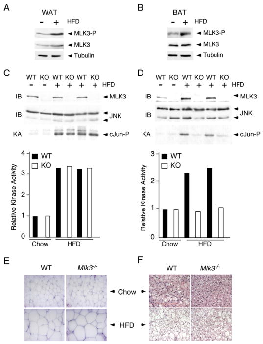
Figure 5. FFA causes MLK3 and JNK activation.

A, B) Wild-type mice were maintained (16 weeks) on a standard diet or on a high fat diet (HFD). MLK3 expression, MLK3 T-loop phosphorylation (Thr-277 and Ser-281), and Tubulin expression in white (epididymal) fat (WAT) and brown (interscapular) fat (BAT) was examined by immunoblot analysis.
C,D) White adipose tissue (C) and brown adipose tissue (D) of wild-type mice (WT) and Mlk3−/−mice (KO) maintained (16 weeks) on a standard diet (chow) and on a high fat diet (HFD) was examined by immunoblot analysis (IB) using antibodies to JNK and MLK3. JNK activity was measured in a kinase assay (KA) using [γ-32P]ATP and cJun as substrates.
E,F) Representative histological sections of white adipose tissue (E) and brown adipose tissue (F) stained with hematoxylin and eosin from wild-type and Mlk3−/− mice fed a standard or high fat diet for 16 wk.
