
Fig. (5).
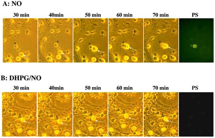
mGluRI activation selectively protects individual neurons from microglial engulfment through maintenance of cellular membrane asymmetry. (A) Microglia and neurons were obtained from the same animal pup litter and were subsequently co-cultured in a ratio of 1:5 (microglia: neurons). Twelve hours after NO (NOC-9, 300 μM) exposure, individual cells were examined over the time points indicated using transmitted light (panels 1-5 in A and B) and fluorescence light with 490 nm excitation and 585 nm emission wavelengths for PS (panel 6 in A and B) of the same microscopy field. A microglial cell is designated by the white arrow and a neuronal cell is identified by a black asterisk. Representative images from 30 minutes (min) to 70 minutes illustrate that NO leads to the specific “tagging” of a neuron with PS externalization and the subsequent engulfment of this cell by a microglial cell. In contrast, application of DHPG (750 μM) 1 hour prior to NO application prevents neuronal PS exposure and the engulfment by microglia.