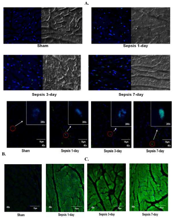
Figure 7.

Immunohistochemistry performed in paraffinized LV tissue sections and visualized by using confocal microscopy. A. Representative photomicrographs (magnification 63x) of LV tissue sections from sham, 1-, 3- and 7-day post-sepsis stained for DNA fragmentation using APO-BrdU TUNEL kit (Invitrogen). TUNEL positive cells are stained green (Alexa Flour 488) in blue nuclei (TO-PRO) along with NOMARSKY DIC (Differential Interference Contrast) images of LV tissue sections.
B. Representative pictomicrographs (magnification 40x) of LV tissues from sham, 1-, 3- and 7-days post-sepsis stained for caspase-3 expression using FITC-conjugated caspase-3 antibody. The cytosol of the LV tissue is stained green (FITC) and the nuclei are counterstained blue with TO-PRO.
C. Representative pictomicrographs (magnifications 200x and 40x) of LV tissues from sham, 1-, 3- and 7-days post-sepsis stained for PARP expression using FITCconjugated caspase-3 antibody. The nucleus of the LV tissue exhibits green (FITC) fluorescence depicting DNA breaks in the blue nuclei which are counterstained with TO-PRO.
