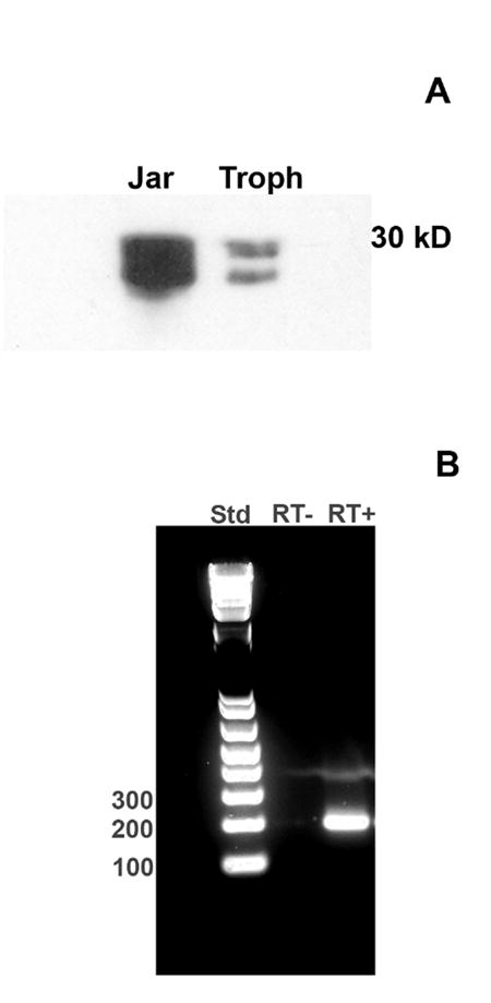
Fig. 3. Analysis of MUC1 expression in trophoblasts by Western blot and RT-PCR.

(A) Trophoblast lysates and human Jar choriocarcinoma cell lysates were examined by Western blot using the CT2 anti-MUC1 antibody (see Methods). A representative blot is shown. (B) Total RNA was extracted from macaque trophoblast cultures and reverse transcribed. The resulting cDNA was used as a template for PCR using primers directed against MUC1 as described in Methods. PCR products were analyzed on 2% agarose gels. DNA standards (Std; left lane) were included. The middle lane shows that no band was produced in the absence of reverse transcription (RT-). The right lane (RT+) shows a prominent band of the predicted size (207 bp). The band was excised from the gel and sequenced. The sequence data confirmed identity with MUC1.
