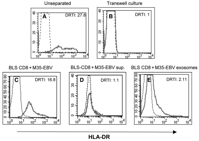
Fig. 5.

Direct cell–cell contact is required for MHC class II transfer. BLS-CD8 T cells and M-35 EBV cells (1:1) were co-cultured mixed (A) or separated by a semi-permeable membrane (0.4 μm) in a transwell dish (B) for 3 h. In a separate experiment, BLS-CD8 Tcells were co-cultured for 24 h with either M35-EBV cells (C), supernatant from a 2 day old M35-EBV cell culture (D) or exosomes purified from M35-EBV cells. The HLA-DR expression on CD8+ gated BLS-CD8 T cells was measured by flow cytometry and analyzed as described in the legend to Fig. 1, and the DRTI is indicated. Thin lines represent HLA-DR staining of the control BLS-CD8 T cells (not incubated with APC, supernatant or exosomes). These results are representative of three independent experiments.
