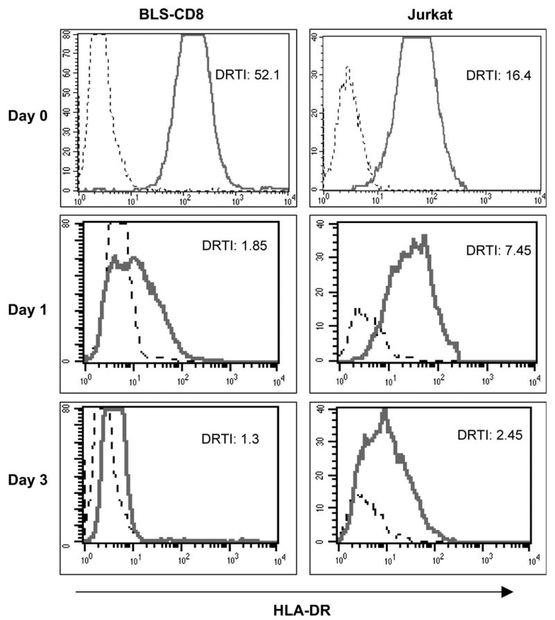
Fig. 6.
Kinetics of MHC class II transfer. BLS-CD8 (left panels) or Jurkat T cells (right panels) were incubated overnight with CFSE-labeled M35-EBV cells. Next day, the CFSE negative T cells were separated by flow sorting, placed on fresh medium and analyzed immediately (Day 0) and at various time points (Days 1 and 3) for HLA-DR expression by flow cytometry.

