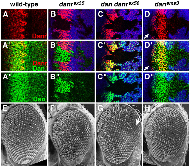
Fig. 3.

Loss-of-function mutants for dan/danr have small, rough eyes. (A) Close-up of the MF from a wild-type eye–antenna disc. (B–D) Discs containing loss-of-function dan/danr clones. Dan (green), Danr (red) and clonal marker (blue) are shown. Clones are marked by the absence of blue staining (see Materials and methods), except in panel B″, where the clonal marker has been removed for clarity. Overlay between red and blue, green and blue, and red and green appears pink, turquoise and yellow, respectively. (B) danrex35 clones do not express Danr, but express higher levels of Dan. (C) dan danrex56 clones do not express either Danr or Dan. (D) danems3 clones express normal levels of Dan; Danr expression is lost from clones anterior to the MF (arrows). (E) SEM picture of a wild-type eye. Eyes from eyFLP;FRT82, arm-lacZ,M/FRT82,danrex35 (F) or eyFLP;FRT82,arm-lacZ,M/FRT82,dan danrex56 (G) individuals. Most of both eyes consist of homozygous mutant tissue (see Materials and methods). danrex35 and dan danrex56 eyes are small and rough. (H) Eye from a danems3 homozygote is slightly small.
