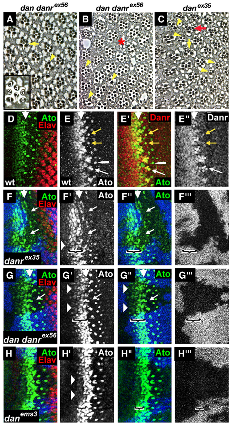
Fig. 4.

danrex35 and dan danrex56 homozygous eye tissues show defects in ommatidial spacing and photoreceptor recruitment. (A–C) Cross sections of eyes from eyFLP;FRT82,arm-lacZ,M/FRT82,dan danrex56 or eyFLP;FRT82,arm-lacZ,M/FRT82,danrex35 flies. Inset: an enlarged wild-type ommatidium showing the positions of the R1–R7 photoreceptors. The internal structure of dan/danr adult mutant eye tissue is variable, ranging from most ommatidia appearing normal (A) to eyes with more frequent defects (B or C) including a loss of photoreceptors (yellow arrowheads), extra small-rhabdomere photoreceptors (C—yellow vertical arrow), defects in rotation (C—red arrow) or gaps in the ommatidial arrangement (B—red asterisk). (D–G) Third instar eye discs. MF is marked by a white arrowhead. (D) Wild-type eye disc double labeled for Elav (red) and Ato (green). (E) Wild-type eye disc double labeled for Ato (green) and Danr (red). Ato and strong levels of Danr expression co-localize in the intermediate groups, in the R8 equivalence group (white arrow) and for a brief period in newly emerged R8 cells (yellow arrows). Shortly afterwards, the strong levels of Danr expression in R8 fade (white long arrowhead). (F–H) Loss-of-function dan/danr clones marked by absence of the blue staining. (F, G) In danrex35 and dan danrex56 mutant clones the stripe of Ato expression (green) anterior to the MF is reduced (horizontal arrowheads), and the number and spacing of the Ato positive R8 cells are altered (arrows). (H) In danems3 mutant clones a narrower stripe of cells ahead of the MF shows a reduction in Ato expression (horizontal arrowheads). The brackets in panels F–H delineate the width of the region ahead of the MF in which Ato expression is reduced in the clones.
