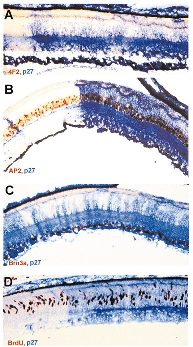
Figure 5.

Various types of nonphotoreceptor neurons in an E12 retina and BrdU+ cells in an E9 retina partially infected with RCAS-cNSCL2. (A) A single layer of horizontal cells (4F2+/LIM+, in red) both in infected (blue) and uninfected regions. (B) Amacrine cells, marked by AP2 immunoreactivity (red), in infected (blue) and uninfected regions. (C) Ganglion cells, identified with Brn3a expression (red or dark), in infected and uninfected regions. (D) BrdU+ cells (red) in infected and uninfected regions in an E9 retina. Magnifications, ×50.
